Infrastructure
The Department of Radiology at SGRRIMHS, Dehradun is a state of art, fully equipped facility providing professional diagnostic and consultative services to referring physicians and patients. The department of Radiology is the premier facility in the state of Uttarakhand. The advance technology & expertise of faculty helps in comprehensive patient evaluation and reaching accurate diagnosis for all the clinical problems of patient's. The department assures the patients that they are receiving some of the finest imaging services available anywhere.
Services Offered
A full range of screening, diagnostic and interventional procedures are performed. A highly trained and professional radiology team performs and interprets these imaging tests.
Department Facilities
Magnetic Resonance Imaging:
The department offers advanced Magnetic Resonance Imaging (MRI) services, where we integrate state-of-the-art technology with exceptional capabilities to deliver precise diagnostic imaging solutions. We are pleased to introduce our latest addition, the 3 Tesla Siemens Magnetom Vida, alongside our esteemed 1.5 Tesla Siemens Avanto SQ engine featuring TIM technology.
3 Tesla Siemens Magnetom Vida:
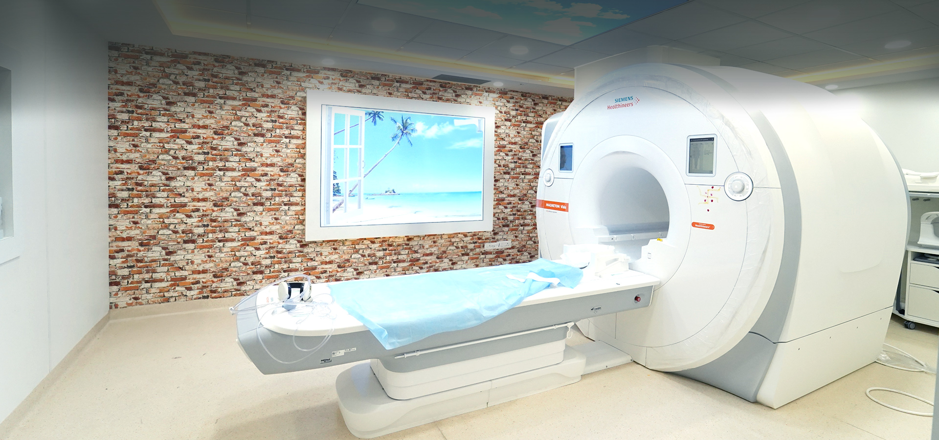
Operating at an ultra-high field strength of 3 Tesla, our MAGNETOM Vida system offers unparalleled clarity and detail for precise anatomical visualization. Groundbreaking BioMatrix technology enables tailored examinations that adapt dynamically to individual patient physiology, ensuring optimal image quality and comfort. Equipped with powerful Tim 4G and Dot engines, it delivers exceptional image quality and workflow optimization for our radiologists. Features include:
- Patient-Centric Comfort and Safety: Our MRI environment prioritizes patient well-being, featuring a spacious gantry room designed to minimize discomfort and acoustic noise, enhancing the overall imaging experience.
- Expanded Patient Eligibility: Inline Compressed Sensing enables free-breathing MRI exams for patients previously unable to undergo traditional breath-hold exams, including those with arrhythmia or limited breath-hold capability.
- Dynamic Imaging Techniques: Compressed Sensing GRASP-VIBE technology facilitates dynamic contrast-enhanced imaging without multiple breath-holds, ensuring diagnostic quality even in challenging cases.
- Whole-Body Imaging: With the Whole-Body Dot Engine and BioMatrix technology, comprehensive oncological scans are performed with reproducible results in minimal time, including diffusion-weighted imaging (DWI) with individual slice adjustment for optimal quality and reproducibility.
- Neurological Imaging: BioMatrix Head/Neck coils provide ultra-fast, high signal-to-noise ratio (SNR) imaging, allowing for precise diagnosis and treatment planning, while advanced techniques such as RESOLVE ensure sharp, high-resolution diffusion imaging.
- Orthopedic Imaging: Ultra-high-density coils maximize SNR and anatomical coverage for accurate diagnosis, with advanced WARP technology reducing image distortions, particularly beneficial for assessing tissue surrounding orthopedic implants.
- Cardiac Imaging: Compressed Sensing Cardiac Cine and PSIR HeartFreeze technologies enable free-breathing cardiac MRI exams with exceptional image quality, expanding eligibility for cardiac imaging.
- Peripheral Angiography: High-resolution, non-contrast and contrast-enhanced angiographic techniques provide detailed vascular imaging for diagnosis and treatment planning.
- Cutting-edge Iron Quantification Technology: Innovative methodologies allow precise measurement and characterization of tissue iron content, crucial in diagnosing and managing iron-related disorders.
- Advanced Motion Correction Software: Ensuring optimal image quality, even in challenging clinical scenarios, enhances diagnostic accuracy and reliability.
1.5 Tesla Siemens Avanto SQ Engine with TIM Technology
The Avanto SQ engine with TIM technology delivers exceptional image quality, equipped with advanced functionalities such as motion correction software and Susceptibility Weighted Imaging (SWI).
Customized Coil Options: Offering comprehensive coil configurations tailored to specific imaging requirements, ensuring optimal results across various clinical scenarios.
Enhanced Patient Experience: With its compact design and in-room amenities including anesthesia and sedation options, our MRI suite fosters patient comfort and convenience.
Dedicated Professional Expertise
Backed by a team of highly skilled professionals committed to delivering precise and reliable diagnostic results, we ensure the highest standard of care for every patient.
Following MRI scans are available:
- Head (Brain), Epilepsy protocol, Diffusion imaging, functional imaging, Tractography, Perfusion studies.
- PNS
- Orbits
- Inner ear
- Neck
- CV junction
- Thorax
- Breast
- Cardiac imaging
- Abdomen
- Pelvis
- Spine
- Spinal cord
- Pituitary
- Extremity bones
- Soft tissues
- Angiography of head, neck, renal, peripheral and other vessels
- Cisternography
- Prostate
- Brachial plexus
- Functional MRI
- Cine dynamic CSF flow study
- Urography
- TM joint
- Neurography
- Myelography
- Iron quantification
- Enterography
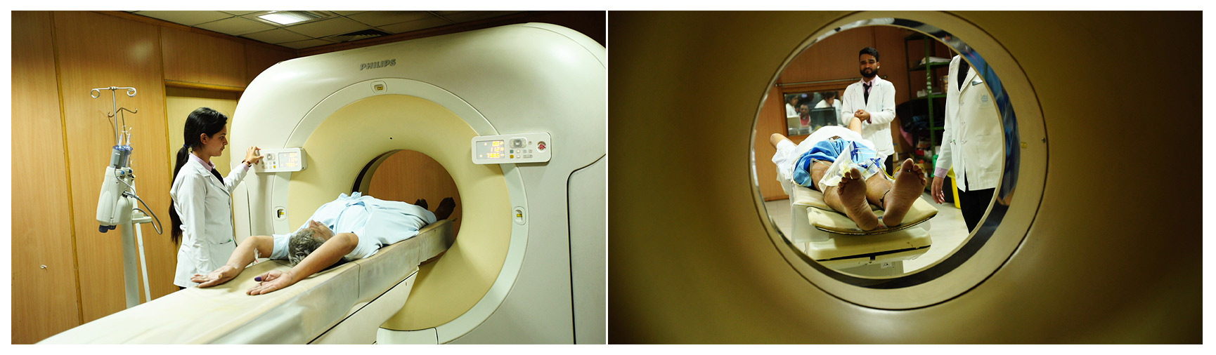
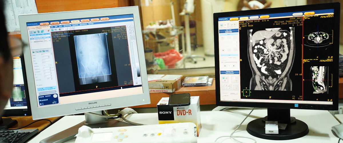
Computed Tomography:
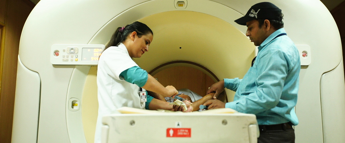
CT scan machine: Computed Tomography (CT) is a method of body imaging in which a thin X-ray beam rotates around the patient. Small detectors interpret the X-rays that make it through the patient or particular area of interest. A computer analyzes the data to construct a cross-sectional image. These images can be stored, viewed on a monitor, or printed on film. CT 128 Philips Incisive Ingenuity is installed which is the state of art facility and one of its kind in state of Uttarakhand that scan the patient in minimum time and with minimum radiation dose. Philips CT 3500 is installed in the emergency block. System is equipped with multi-projection volume reconstruction, 3D software- surface and volumetric renderings. The following CT studies are performed.
- Head. Non contrast and contrast enhanced scans are done. Also perfusion studies are done to assess the flow in diseased and normal brain tissue.
- PNS
- Neck
- Chest
- Abdomen
- Multiphasic studies of liver
- Spine
- Bones & joints
- Soft tissues
- Cisternography
- Coronary Angiography
- Angiography –cerebral, thoracic & abdominal aorta, Renal, Carotid, upper and lower limb arteries etc.
- Virtual bronchoscopy, colonoscopy, endoscopy etc
- Thin 0.6 mm slices for inner ear with excellent 3D reconstructions.
- 3D of bones and joints etc.
- CT Guided Biopsy of lung mass, liver mass, renal mass, bone tumors and other lesions.
Digital Subtraction Angiography:
DSA: The Department possesses a high end Allenger Altima ADV Machine for Vascular Interventions.
We offer full range of Coronary and Peripheral Diagnostic and Interventional Procedures. Vascular services for all patients encompass diagnostic Angiography, Stents, Thrombolysis, Inferior Vena cava (IVC) filter placement, Management of Deep Venous Thrombosis (DVT), Hemodialysis grafts, Embolization of Vascular lesions and Venous Access. In addition Neuroangiography and Interventions facilities are available which include Endo-vascular Aneurysmal coiling, AVM and Tumor Embolizations and Thrombolysis for Stroke patients.
Mammography:
Mammography: The department is equipped with the latest GE RT mammography machine with special viewing monitors for the radiologist. This is coupled with breast ultrasound which typically improves mammographic and clinical skills.
Breast cancer is the second biggest killer of women next to cervical cancer.
A base line mammogram is recommended after the age of 40 years with regular yearly screening after 40 years of age, especially more so in those with family history of disease. Regular mammography screening helps detect cancer early and thus save many precious lives. It is also advisable prior to HRT (Hormone replacement therapy), in any skin changes, nipple discharge, persistent pain, lump etc. Mammograms and ultra-sound scans are complimentary investigations and help detect lesions that may not be apparent in one investigation. Mammography is however highly sensitive in fatty breasts and is the recommended investigation for screening.
We have latest Philips machine (IU- 22) for sonomammography. It also has latest facility of breast elastography which is very helpful in categorizing breast lesions as benign and malignant. There is a dedicated breast MRI with special breast coils to perform dynamic breast MRI examination.
Thus for breast imaging we have
-Mammography
-latest GE equipment.
-Breast ultrasound (sonomammography).
-Breast elastography. -Breast MRI
Digital Radiography:
The department has installed Digital Radiography System by Allengers Medical System MARS 50 Digital Radiography -630mA, 150 kV & ASTRO65 DR 800mA, 150 kV
Advantage of Digital Radiography (DR): Digital image processing (overcome errors in exposure factor selection) for enhanced details.
- Preview image is available in less than 3 seconds (no long wait for patients).
- Perfectly dry film printing (no chemicals).
- No physical movement of cassettes (faster operation).
- Wide dynamic range (soft tissues and hard bones can be seen together).
- Superb image quality due to fine pixels and high contrast image.
- Post processing adds more to the image quality.
- Efficient and user friendly work flow due to DICOM based system.
CR Radiography:
CR Radiography plus Fluoroscopy System: Real-time dynamic imaging using x-rays (for the purpose of procedural guidance) is referred to as "fluoroscopy". User-friendly, high-performance, CR based x-ray system is available in the department capable of producing good-quality x-ray images with reasonable use of radiation dose. Such imaging techniques are commonly associated with contrast medium aided examinations of the gastrointestinal tract.
Following X-ray Special procedures are performed in the department:
- Barium Procedures like Ba Swallow, Ba Meal, Ba Meal FT and Ba Enema
- MCU
- Urethrogram
- IVU
- HSG
The following Radiography, Digital Radiography and CR System machines are in the department
- GE HF Advantage / Simplex - 400 mA
- EPSILON – ASTRO 65 DR - 800 mA
- Allengers HF Medical System MARS 50 - 600 mA
- Allengers Medical Sysm 50 Plus Fluoroscopy- 800 mA
- EPSILON EPCORSA - 40 - 600 mA
- Allengers 325mA
- Allengers Mars 50 – 630 mA
- ADONIS Fixed X – Ray - 630 mA
CR system
- Fuji CR System-3
- Fuji Dry pix 7000-2
- Fuji Dry pix 7000
For mobile radiography, ten portable X-ray machines are available.
Ultrasound and Colour Doppler:
The department of radiology are equipped with following Ultrasound machines which contribute to the high level of confidence of the Radiologist for evaluation of the lesions and serve as excellent guiding tools for the various Diagnostic and Therapeutic procedures.
Machines in the Department of Radiology
- Mindray Resona 19
- Philips Affiniti 30
- Samsung Accuvix-XG
- Toshiba Nemio-XG SSA-580A
- Mindray DC-30
- Mindray DC-30
- Mindray DC-60
- GE Voluson E8
- GE Voluson S8
Machines in Hospital Premises
- Philips CX-50
- GE VIVID E-9 4D
- SONOSITE MICROMAXX
- L & T SONATA PLUS
- Toshiba SSA-320A
- MIND RAY DP-1100 Plus
- SONOSITE M-TURBO
- Toshiba SSA550A
- Toshiba SSA-510A
- MINDRAY DC-70S
- Hitachi ALOKA – F31
- NIDEK Echo Scan US 4000
- Philips Portable Cx-50
- Philips Affinity CVX
- Fujifilm Sonosite M-Turbo
These machines are used for the diagnostic purpose to carry out various ultrasound examinations of the abdomen, pelvis and small parts, as well as intracavitary (transrectal and transvaginal) examinations, antenatal scan, fetal doppler and anomaly scan. The department has an advanced USG scanner with elastography and 3D and 4D applications.
The tissue Harmonics helps for high resolution at depth and the Cross Beam Imaging facility makes the lesion to stand out with better border delineation.
The 3D/ 4D Application further helps to understand better the relations of the lesion with organs in the vicinity.
The Color Doppler scanners are extensively used to assess the vascularity of the tumors. The arterial and venous Doppler examinations are carried out for the evaluation of patients with associated peripheral Vascular Diseases and screening as well as diagnosis of the Deep Venous Thrombosis.
The Ultrasound Equipment in the Interventional Radiology Unit is an integral part of the set up as it serves as an important guide for the Vascular as well as Non-vascular procedures for access and precise placement of the needles.
The portable Ultrasound unit delivers the conveniences and versatility with its application in:
- The ICU for Emergency Scanning of ICU patients
- Bedside Drainage procedures like
: – Pleural & Peritoneal Tapping
: – Abscess Aspiration
: – Indwelling Pigtail Drainage
- Nerve Block
Being a teaching institute, the department has regular teaching for radiologists and staff memberswith internal auditing and professional appraisal from time to time. The department also has ultramodern library, museum and seminar rooms.. The PG courses in the department have startedfrom 2013.
Subspeciality services.
Musculoskeletal Radiology (Ultrasonography, MRI, and Intervention):
We specialize in Musculoskeletal Radiology, offering a comprehensive array of diagnostic and interventional services for conditions affecting the muscles, joints, tendons, ligaments, and soft tissues. Our advanced imaging techniques include MRI and ultrasound, providing detailed assessments of musculoskeletal structures to diagnose injuries, inflammation, tears, and degenerative changes. Additionally, our skilled team performs interventional procedures such as guided injections, aspirations, and biopsies, ensuring accurate diagnosis and effective treatment planning for musculoskeletal conditions.
Breast Radiology (Mammography, Ultrasonography, MRI, and Intervention):
Our radiology department excels in Breast Radiology, offering a comprehensive range of services including Mammography, Ultrasonography, MRI, and Interventional procedures. Mammography is a vital screening tool for detecting breast cancer in its early stages, while Ultrasonography provides detailed images to further evaluate abnormalities found on mammograms or clinical exams. MRI offers high-resolution imaging, particularly useful for assessing breast tissue in certain cases. Additionally, our skilled team performs Interventional procedures such as biopsies and aspirations, providing accurate diagnosis and treatment guidance for breast-related conditions.
Various intervention procedures performed in our department:
HEPATOBILIARY INTERVENTIONS
- Hepatocellular carcinoma treatment by:
- Transarterial embolisation using PVA particles
- Transarterial chemoembolisation using doxorubicin Lipiodol/DEB/DC beads
- Transarterial chemoinfusion
- Radiofrequency ablation
- Transarterial radioembolisation
- Cholangiocarcinoma Treatment:
- Hepatic metastases:
- Radiofrequency ablation depending on indication
- Transarterial chemoembolisation
- Obstructive biliopathy:
- Percutaneous transhepatic biliary drainage with biliary Stenting
- Percutaneous biliary drainage with balloon plasty for benign biliary stricture
- Percutanteous calculus extraction using Stone Extraction Basket.
- Portal hypertension management
- Transhepatic intrajuglar porto systemic shunt
- Balloon occluded retrograde transvenous obliteration for gastric varices(antegrade and retrograde)
- Hepatic venoplasty for management of budd chiari syndrome
- Portal vein embolisation as adjunct to surgery
- Hepatic arterial bleeding/aneurysmal coiling/glue
- Hepatic venous pressure gradient to evaluate portal hypertension
- Transjuglar liver biopsy
VASCULAR MALFORMATION:
- Arterio-venous malformation treatment using sclerosant/coils
- Venous malformation sclerotherapy
- Pulmonary AVF using vascular plug/coils
- Lymphatic malformation
UROGENITAL:
- Renal tumor radiofrequency ablation
- Non resectable renal tumor – embolisation
- Renal AVM embolisation
- Renal artery stenosis – angioplasty and stenting
- Percutaneous nephrostomy
- For assisting in endoscopic lithotomy
- For drainage in pyonephrosis/obstructed systems
- With antegrade stenting in malignant etiology
- Percutaneous cystosomy(under local anaesthesia without any surgical scar
- Pelvic congestion syndrome (ovarian vein emolisation)
- Uterine fibroid embolisation
GASTROINTESTINAL:
- Endovascular management of post operative bleeders
- Vascular stenting(SMA/Celiac/Renal artery)
- Percutaneous gastrostomy with or withour gastropexy under local anaesthesia
- Endovenous
- Nasojejunal tube(freka)insertion (under Fluoroscopy)
- Esophageal/Gastro-duodenal stenting in cases where endoscopy is not feasible or available
VENOUS:
- Superior venae caval/IVC stenting
- Subclavian vein stenting in malignant breast carcinoma or other
- Tunneled venous catheter (Chemoport/Hickmann/Dialysis Catheter)
- Central line/dialysis catheter insertion under USG guidance with proper positioning into SVC-RA junction under C-ARM
- Limb vessel thrombolysis
- IVC filter placement
Arterial procedure:
- Peripheral DSA & angioplasty
- Peripheral thrombolysis
Other procedures:
- Cholecystostomy
- USG/TRUS/TVS guided biopsies
- CT guided biopsies
- Painless percutaneous catheter insertions for liver abscess/abdominal peipancreatic collections/empyema with or without thoracocentesis/ ascitic or pleural effusion drainage using co axial techniques
- Tunneled ascitic fluid catheter(for long term home ascitic drainage)
NEUROINTERVENTIONS:
Neuroangiography and Interventions facilities are available which include Endo-vascular Aneurysmal coiling, AVM and Tumor Embolizations and Thrombolysis for Stroke patients.
MUSCULO SKELATAL SYSTEM:
- Radiofrequency ablation for osteoid osteoma
- Muskuloskeletal/bone biopsies
- Joint injections for tendinitis, bursitis
- Vertebroplasty
LYMPHATIC INTERVENTIONS:
- Lymphangiography using lipidol
- Post operative lymphocele
- Lymphatic malformation
Varicose vein ablation using radiofrequency and laser Pain management using alcohol / Radiofrequency
- Coeliac plexus block
- Lumbar sympatholysis etc
Classroom teaching equipment
Newline Smartboard TT 5521Q a state-of-the-art interactive touchscreen with high-resolution visuals, multi-user capability, built-in Android system, and wireless connectivity, it offers seamless collaboration and versatile functionality. Durable and compatible with various devices and software, engaging presentations and interactive learning.
Publications from the department:
- Sarda P, Sharma R, Mishra S. Real-World Outcomes of Intravascular Lithotripsy in Calcified Below-the-Knee Arteries. Journal of Contemporary Clinical Practice. 2025 April 11(4) 8 – 13.
- Chauhan T, Sharma R, Sharma VK. Ultrasonographic Assessment of Placental Thickness and Its Correlation with Neonatal Birth Weight. Int J Pharm Clin Res. 2024;16(3):351-8.
- Arora M, Lakhera D, Rawat K, Thakker V, Randhawa LS, Patel A, Agarwal R, Azad RK.Diffusion-weighted Imaging: New Paradigm in Diagnosis of Early Acute Pancreatitis.Annals of African Medicine.:10-4103.
- Arora M, Abhishek A, Singh N, Thakker V, Azad S, Azad R, et al. Prediction ofPathological Risk Stratification using Computed Tomography Features in GastrointestinalStromal Tumors: A Retrospective Observational Study. Journal of Clinical and DiagnosticResearch 2024 18(3). ISSN-2249-782X.
- Randhawa LS, Semwal A, Srivastava RK, Hernot S, Azad RK, Kaintura M, et al. ADetailed Assessment of Variations of Ethmoid Roof, Olfactory Fossa, and AnteriorEthmoidal Artery on CT Scan of Paranasal Sinuses of 200 Patients. Indian Journal ofOtolaryngology and Head & Neck Surgery 2023 July ; 75(4) :23-30. ISSN-2231-3796
- Arora M, Randhawa LS, Lakhera D, Thakker VD, Abhishek KA, Singh, D, et al. Inter- observer Reproductibility of RENAL Nephrometry score in comparison to simplifiedPADUA Renal (SPARE) Nephrometry Score. ‘Urology Annals’. 2023 8 ;11(11). ISSN-0974-7796.
- Arora M, Waikhom PD, Azad S, Thakker V, Azad RK, Srivastava R,. Impact of COVID-19 on the education and mental well-being of postgraduate medical students: CrossSectional Survey from an Indian Medical College. Journal of Education and HealthPromotion 2022 28;11:294. ISSN-2277-9531.
- Thakker V, Sarda P, Ruhela V, Arora M, Sharma R, Azad RK. Role of endovasculartreatment in dysfunctional hemodialysis fistulae: A single center experience. IndianJournal of Nephrology. July 29, 2022. 32(5). ISSN-0971-4065.
- Sharma R, Thakker V, Sharma RB, Arora M, Baisoya A, Azad R. MultidetectorComputed Tomography Angiographic Evaluation of Anatomical Variations in PoplitealArtery Branching: A Retrospective Study from Northern India. Journal of Clinical &Diagnostic Research. 2022 Jul 1;16(7). ISSN-2249-782X.
- Sharma R, Thakker V, Sharma RB, Arora M, Sarda P, Ahuja M, Randhawa LS, Azad RK.Effect of vaccination on the HRCT profile of COVID-19 patients–A single-centerexperience. Journal of Family Medicine and Primary Care. 2022 Jun 1;11(6):2938-44.ISSN-2249-4863.
- Sarda P, Thakker V, Waikhom PD, Arora M, Sharma R, Baisoya S, Rawat J, Azad RK.Pneumomediastinum: Radiological profile and associations of uncommon complication ofCOVID-19. Journal of Family Medicine and Primary Care. 2022 Feb;11(2):537. ISSN-2249-4863.
- Lakhera D, Sarda P, Waikhom P, Bhuyan D, Duara BK. Role of Non-Invasive Imaging inCharacterization of Soft Tissue Vascular Anomalies: Comparison of Ultrasound withContrast-Enhanced Magnetic Resonance Imaging. J Clin Diagn Res 2020;14:01-05
- Lakhera D, Azad RK, Azad S, Singh R, Sharma R. Magnetic Resonance ImagingCerebrospinal Fluid Hydrodynamics in patients with meningitis. Journal of clinicalimaging science 2020; 10:29. ISSN – 2156-7514.
- Juneja A, Azad R, Malhotra A. Magnetic Resonance Imaging Evaluation of CerebralMicrobleeds: A comparative Analysis of Susceptibility Weighted Imaging and T2Gradient Recalled Echo Sequences.. J Clin Diagn Res 2020; 14:6-12. ISSN-2249-782X.
- Arora P, Rawat K, Azad R, Chouhan K. Assessment of Cerebrospinal FluidHydrodynamics using MRI in post craniospinal surgery patients.. Indian Journal ofRadiology and Imaging. 2021 Apr; 31(2): 304-310. ISSN-0971-3026• Malhotra A, Azad R, Srivastava R, Juneja A, Lakhera D, Chauhan T. Assessment ofMultidetector computed tomography signs of unilateral vocal cord palsy: Do we reallyneed to evaluate coronal reformatted images ? . J Clin Diagn Res 2017; 11:19-23. ISSN -2249-782X.
- Azad R, Tayal M, Azad S, Sharma G, Srivastava R. Qualitative and quantitativecomparison of contrast enhanced fluid attenuated inversion recovery, magnetization transfer, spin echo, and fat saturation T1-Weighted Sequences in Infectious Meningitis..Korean J Radiol 2017; 18:973-982. ISSN-1229-6929.
- Azad R, Mittal P, Malhotra A, Gangrade S. Detection and Differentiation of focalintracranial calcifications and chronic microbleeds using MRI.. J Clin Diagn Res 2017;11:19-23. ISSN-0973-709X.
- Ahmad A, Azad S, Azad R (Cos Author). Differentiation of leptomeningeal and vascularenhancement on post contrast FLAIR MRI sequence: Role in early detection of infectiousmeningitis., J Clin Diagn Res 2015; 9:8-12. ISSN-0973-709X.
- Aggarwal A, Azad R (Cos Author), Ahmed A, Gupta P, Arora P. Additional merits ofsingle thick slab two dimensional MR Myelography in spinal imaging.. J Clin ImagingSci, Dec 2012; 2:84. ISSN – 2156- 7514.
- Azad R, Azad S, Shukla AK, Arora P. Role of screening of whole spine with sagittal MRIwith MR Myelography in early detection and management of occult intrasacralmeningocele.. Asian J Neurosurg 2012; 7:162-6. ISSN -1793 -5482.
- Azad R, Magu S, Gathwala G. Sonographic Assessment of Thymus and Measurement ofThymic. Size in healthy Neonates from North India.. JCDR, 2011; 5:1406-9. ISSN-0973-709X.
- Kamra P, Azad R, Prasad KM, Jha S, Pradhan S, Gupta RK. Infectious meningitis:Prospective evaluation with Magnetisation transfer-MR imaging. BJR 2004; 77: 387-94.ISSN -0007-1285.(Part of this work has been presented at European Congress ofRadiology, April 1, 2002).
- Azad R, Gupta RK, Kumar S, Pandey CM, Prasad KM, Hussain N, Hussain M. IsNeurocysticercosis a risk factor in coexistent intracranial disease- MRI based study?.JNNP 2003; 74: 359-61. ISSN-0022-3050